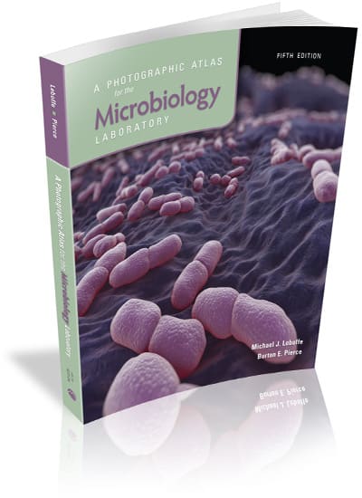
A Photographic Atlas for the Microbiology Laboratory, 5e

By Michael J. Leboffe, Burton E. Pierce  •  
 2021  
•  352
 Pages
Loose-leaf
Format • 
Print ISBN  9781617319037
• eBook ISBN  9781617319068
Suggested Student Retail Price

$48.40

Purchase
Request review copy
eBook Options

This full-color atlas is intended as a visual reference to supplement laboratory manuals or instructor-authored exercises for introductory microbiology laboratory courses. The atlas can be used alone but also has been designed to be used in conjunction with Exercises for the Microbiology Laboratory, 5e, by Leboffe & Pierce, with images keyed to specific exercises.

A Photographic Atlas for the Microbiology Laboratory, 5e is part of our CustomLab program. For more information on customizing this book contact our CustomLab Editor.

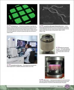
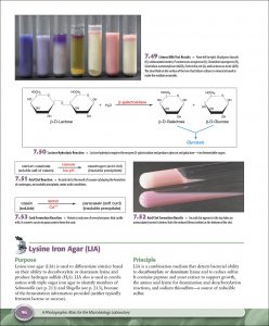
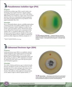
Examples of interior pages:
                       

See More Information
Features
What's New in this Edition
Table of Contents
Versions and Packages
Features
  • More than 700 full-color, high-quality, color-accurate photographs serve as a visual reference for students in the laboratory and aid in interpreting results in the lab.
  • The purpose and principle are set forth for each technique and test in the book so students will understand the “why” as well as the “how” in learning to be proficient microbiologists.
  • A section on microscopy provides practical information about how to operate a microscope.
  • Informative color diagrams, clearly written captions, and additional text provide helpful information to make the atlas easy to use and understand.
What's New in this Edition
  • A significant number of the 700 photographs and light micrographs have been replaced or edited to improve contrast and color.
  • New transmission and scanning electron micrographs have been included to enhance the sections on bacterial structure.
  • Revised organismal sections that take a phylogenetic approach. Relevant taxa are included in the photo captions to assist the reader in maintaining their bearings as they study taxonomy.
  • The printing of bacterial micrographs with the cells on the page approximating their size as seen through a 1000X oil immersion lens.
Table of contents
Section 1 Introduction
Microbial and Eukaryotic Systematics
The Working Microbiologist
A Word About Laboratory Safety
Section 2 Isolation Techniques and Selective Media
Streak Plate Methods of Isolation
Bacteroides Bile Esculin (BBE) Agar
Bismuth Sulfite Glucose Glycine Yeast (BiGGY) Agar
Chocolate II Agar
Columbia CNA with 5% Sheep Blood Agar
Deoxycholate (DOC) Lactose Agar
m Endo Agar LES
Eosin Methylene Blue (EMB) Agar (Levine)
Hektoen Enteric (HE) Agar
MacConkey Agar
Mannitol Salt Agar (MSA)
Phenylethyl Alcohol (PEA) Agar
Pseudomonas Isolation Agar (PIA)
Sabouraud Dextrose Agar (SDA)
Salmonella–Shigella (SS) Agar
Thiosulfate Citrate Bile Sucrose (TCBS) Agar
Vogel and Johnson (VJ) Agar
Xylose Lysine Deoxycholate (XLD) Agar
Section 3 Bacterial Growth
Growth Patterns on Agar
Growth Patterns in Broth
Aerotolerance
Cultivation of Anaerobes—Anaerobic Jar
Cultivation of Anaerobes—Fluid Thioglycollate Broth (FTB)
Cultivation of Anaerobes—Cooked Meat Broth
Section 4 Microscopy
Types of Microscopy
Section 5 Bacterial Cellular Morphology, and Simple and Negative Stains
Simple Stains
Negative Stain
A Gallery of Bacterial Cell Diversity
Section 6 Bacterial Cell Structures and Differential Stains
Gram Stain
Acid-fast Stains
Capsule Stain
Endospore Stain
Flagella Stain
Wet Mount and Hanging Drop Preparations
Miscellaneous Structures
Section 7 Differential Tests and Multiple-Test Systems
Part A: Differential Tests
Bacitracin Susceptibility
β-Lactamase Test
Bile Esculin Agar (Esculinase Test)
Blood Agar
CAMP Test
Casein Hydrolysis (Casease Test)
Catalase Test
Citrate Utilization
Coagulase and Clumping Factor Tests
Decarboxylase Tests
DNA Hydrolysis (DNase Test)
Fermentation Tests (Purple Broth and Phenol Red Broth)
Gelatin Hydrolysis (Gelatinase Test)
Indole Production
Kligler’s Iron Agar
Lipid Hydrolysis (Lipase Test)
Litmus Milk Medium
Lysine Iron Agar (LIA)
Malonate Utilization
Methyl Red Test (MR-VP Broth)
Motility Agar
Nitrate Reduction
Novobiocin Susceptibility
Oxidase Test
Oxidation–Fermentation (O-F) Test
Phenylalanine Deaminase Test
L-Pyrrolidonyl–β-Naphthylamide Hydrolysis (Pyrrolidonyl Arylamidadse [PYR] Test)
Starch Hydrolysis (Amylase Test)
Sulfur Reduction (SIM Medium)
Sulfamethoxazole-Trimethoprim (SXT) Susceptibility Test
Triple Sugar Iron Agar (TSIA)
Urea Hydrolysis (Urease Tests)
Voges-Proskauer Test
Part B: Multiple-Test Systems
API 20 E Identification System for Enterobacteriaceae and Other Gram-Negative Rods
BBL CRYSTAL Identification System—Enteric/Non-Fermenter (E/NF)
EnteroPluri-Test
Vitek 2–Compact
Section 8 Serology
Precipitation Reactions
Agglutination Reactions
Enzyme Immunoassay (EIA)/Enzyme-Linked Immunosorbent Assay (ELISA)
Fluorescent Antibody (FA) Technique
Section 9: Molecular Techniques
DNA Extraction
DNA Sequencing
Polymerase Chain Reaction
Gel Electrophoresis and DNA Hybridization
DNA Hybridization and Microarrays
Matrix-Assisted Laser Desorption/Ionization Time of Flight (MALDI-TOF) Mass Spectrometry (MS)
Section 10 Viruses
Introduction to Viruses
Human Immunodeficiency Virus (HIV)
Viral Cytopathic Effects in Cell Culture
Hemadsorption in Cell Culture
Section 11 Domain Bacteria
Bergey’s Manual of Systematic Bacteriology, 2nd ed., Volume 1
Phylum Aquificae
Phylum Thermotogae
Phylum Deinococcus-Thermus
Phylum Chloroflexi
Phylum Cyanobacteria (Cyanophyta)
Phylum Chlorobi
Bergey’s Manual of Systematic Bacteriology, 2nd ed., Volume 2
Phylum Proteobacteria
Class Gammaproteobacteria (Volume 2B)
Class Alphaproteobacteria (Volume 2C)
Class Betaproteobacteria (Volume 2C)
Class Deltaproteobacteria (Volume 2C)
Class Epsilonproteobacteria (Volume 2C)
Bergey’s Manual of Systematic Bacteriology, 2nd ed., Volume 3
Phylum Firmicutes
Class Bacilli
Class Clostridia
Bergey’s Manual of Systematic Bacteriology, 2nd ed., Volume 4
Phylum Bacteroidetes
Phylum Spirochaetes
Bergey’s Manual of Systematic Bacteriology, 2nd ed., Volume 5
Phylum Actinobacteria
Section 12 Bacterial Pathogens
Aeromonas spp.
Bacillus anthracis
Bacillus cereus
Bacteroides fragilis
Bordetella pertussis
Borrelia burgdorferi
Brucella melitensis
Campylobacter jejuni
Citrobacter spp.
Clostridioides (Clostridium) difficile
Clostridium botulinum
Clostridium perfringens
Clostridium septicum
Clostridium tetani
Corynebacterium diphtheriae
Escherichia coli
Fusobacterium nucleatum and Fusobacterium necrophorum
Haemophilus influenzae
Helicobacter pylori
Klebsiella pneumoniae
Legionella pneumophila
Listeria monocytogenes
Mycobacterium leprae
Mycobacterium tuberculosis
Neisseria gonorrhoeae
Neisseria meningitidis
Nocardia Species
Proteus mirabilis
Pseudomonas aeruginosa
Rickettsia rickettsii
Salmonella enterica subsp. enterica
Shigella dysenteriae
Staphylococcus aureus
Staphylococcus epidermidis
Streptococcus agalactiae
Streptococcus mutans
Streptococcus pneumoniae
Streptococcus pyogenes
Treponema pallidum
Vibrio cholerae
Yersinia pestis
Section 13 Domain Archaea
Crenarchaeota
Euryarchaeota
Nanoarchaeota, Korarchaeota, and Thaumarchaeota
Section 14 Domain Eukarya: Simple Eukaryotes
Group Excavata
Group SAR/HA
Group Archaeplastida
Group Unikonta
Section 15 Fungi of Clinical Importance
Ascomycetous Yeasts
Basidiomycetous Yeasts
Monomorphic Molds
Section 16 Protozoans of Clinical Importance
Group Excavata
Group SAR
Group Unikonta
Section 17 Parasitic Helminths
Trematode Parasites Found in Clinical Specimens
Cestode Parasites Found in Clinical Specimens
Nematode Parasites Found in Clinical Specimens
Section 18 Quantitative Microbiology
Standard Plate Count (Viable Count)
Direct Count (Petroff-Hausser Counting Chamber)
Plaque Assay for Determination of Phage Titer
Urine Streak—Semiquantitative Method
Section 19 Medical, Environmental, and Food Microbiology
Part A: Medical Microbiology
Antimicrobial Susceptibility Tests (Kirby-Bauer Method and E-Test)
Snyder Test
Clinical Sample Collection and Transport
Part B: Environmental Microbiology
Environmental Sampling: The RODAC Plate
Water Quality Testing: Colilert Test
Water Quality Testing: Membrane Filter Technique
Water Quality Testing: Multiple Tube Fermentation Method (Most Probable Number)
Water Quality Testing: Quanti-Tray/2000 Kit
Bioluminescence
Winogradsky Column
Nitrogen Cycle
Sulfur Cycle
Part C: Food Microbiology
Methylene Blue Reductase Test
Yogurt
Section 20 Host Defenses
Differential Blood Cell Count
Immune Cells and Organs
Appendix A: Biochemical Pathways
Oxidation of Glucose: Glycolysis, Entner-Doudoroff, and Pentose Phosphate Pathways
Oxidation of Pyruvate: The Citric Acid Cycle and Fermentation
References
Index
cross-circle